Understanding Sacroiliac Joint Dysfunction
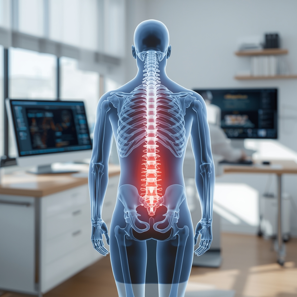
Sacroiliac joint dysfunction (SIJD) refers to pain originating from the sacroiliac joints, which connect the sacrum to the pelvis. This condition can mimic other lower back or hip issues, making accurate diagnosis critical. Symptoms often include localized pain in the lower back or hips, radiating discomfort down the legs, and difficulty with movement. Because SIJD lacks a single definitive test, healthcare providers rely on a combination of clinical evaluation and diagnostic tools. Early and precise identification is essential to guide treatment, as misdiagnosis can lead to ineffective interventions. Chiropractors play a key role in assessing these joints, using their expertise to differentiate SIJD from conditions like lumbar disc herniation or sciatica. Understanding the available diagnostic methods helps patients appreciate the thoroughness of the evaluation process and the importance of collaboration with qualified professionals.
Physical Examination Techniques
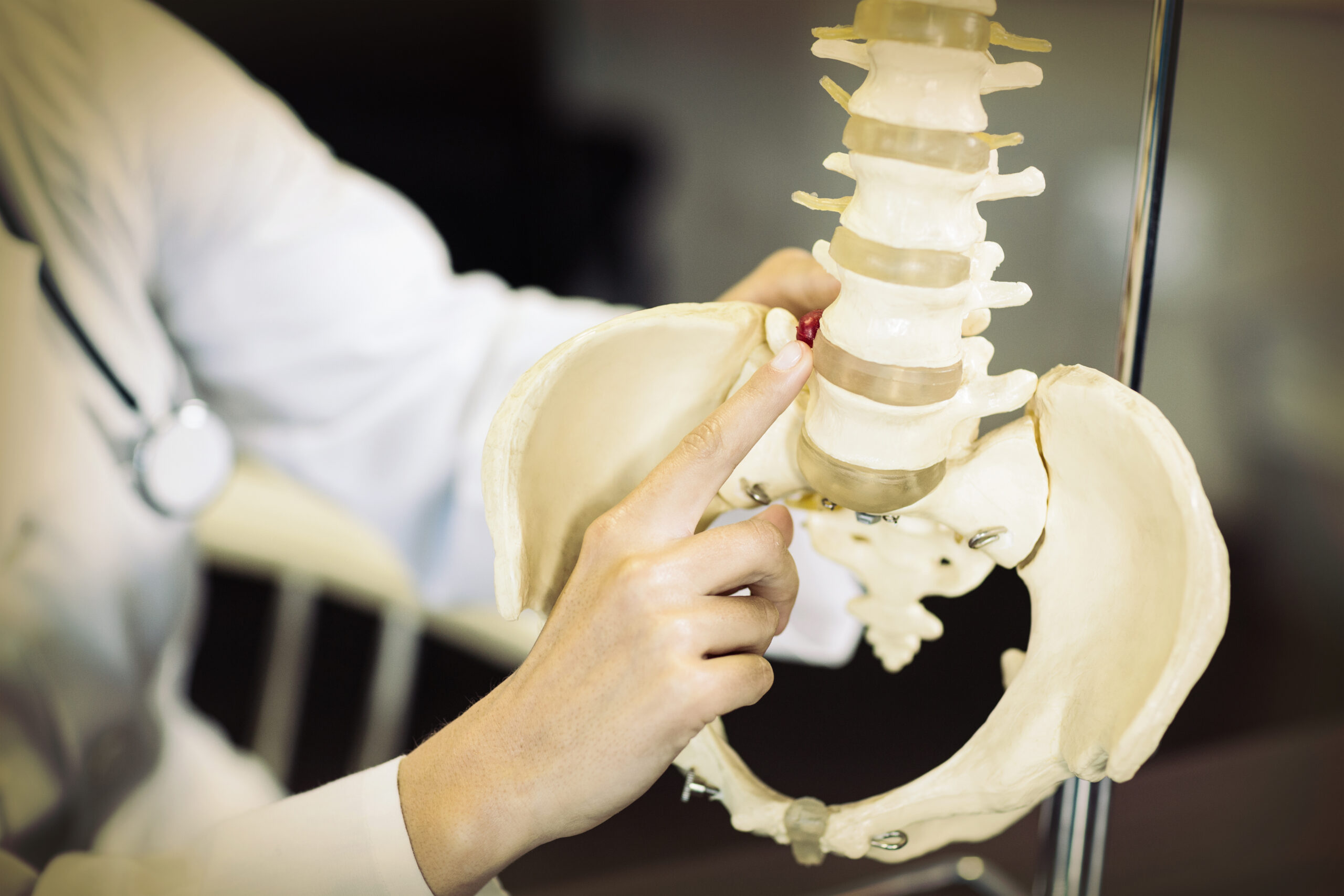
Physical exams form the foundation of diagnosing SIJD. Chiropractors and other providers use specific maneuvers to assess joint mobility, tenderness, and pain patterns. Tests like the FABER (Flexion, Abduction, External Rotation) test or the Gaenslen’s test apply pressure to the sacroiliac joints, reproducing symptoms to confirm dysfunction. Palpation of the iliac crests and sacrum helps identify tenderness or asymmetry. While these methods are non-invasive and cost-effective, their accuracy depends on the provider’s experience. For instance, a study in the Journal of Manipulative and Physiological Therapeutics noted that physical exams alone may miss subtle cases. However, when combined with patient history, they provide valuable initial insights. Patients should understand that these exams are part of a broader diagnostic approach and may be followed by imaging or other tools for confirmation.
Imaging Studies for Diagnostic Clarity
Imaging techniques like X-rays, MRI, and CT scans are commonly used to visualize the sacroiliac joints. X-rays can detect structural abnormalities such as arthritis or fractures but may not capture soft tissue issues. MRI offers detailed views of ligaments, discs, and inflammation, making it useful for identifying conditions like sacroiliitis. CT scans provide high-resolution images of bone structures, aiding in cases of suspected degeneration. However, these tools have limitations: imaging may show abnormalities unrelated to the patient’s symptoms, leading to overdiagnosis. For example, degenerative changes on an MRI are common in asymptomatic individuals. Chiropractors often interpret these findings alongside clinical exams, emphasizing that imaging is a supportive tool rather than a standalone diagnostic method. Patients should discuss the risks and benefits of each test with their provider to avoid unnecessary procedures.
Injections as Diagnostic and Therapeutic Tools
Diagnostic injections, such as sacroiliac joint blocks, are used to confirm SIJD by temporarily numbing the joint. If pain relief occurs after the injection, it suggests the sacroiliac joint is the pain source. These injections also serve a therapeutic role, providing short-term symptom relief. However, they are not without risks, including infection or nerve damage, though these are rare. The accuracy of injections depends on proper technique and interpretation. For instance, a positive response may indicate SIJD, but it could also reflect other issues. Chiropractors often use injections in conjunction with other methods, ensuring a comprehensive approach. Patients should be aware that while injections can be helpful, they are part of a broader diagnostic strategy and may not resolve underlying causes. Always consult a qualified provider to determine if this approach is suitable for your situation.
Emerging Technologies and Alternative Tools
Advancements in technology have introduced new diagnostic options for SIJD. Ultrasound, for example, allows real-time visualization of joint movement and soft tissues, offering a dynamic assessment. While less common than MRI or X-rays, it is non-invasive and avoids radiation exposure. Electromyography (EMG) may help identify nerve involvement, though its role in SIJD is limited. Some practitioners use motion analysis systems to evaluate gait and posture, which can indirectly point to sacroiliac dysfunction. These tools are still evolving, and their accuracy varies. Chiropractors often integrate them with traditional methods, emphasizing that no single tool is foolproof. Patients should discuss the availability and appropriateness of these technologies with their provider, ensuring they align with their specific needs and clinical context.
Integrating Diagnostic Approaches for Accurate Outcomes
Diagnosing SIJD requires a multifaceted approach, combining physical exams, imaging, injections, and emerging technologies. No single method is infallible, so providers rely on clinical judgment to interpret findings. For example, a patient with positive physical exam results and MRI evidence of inflammation may receive a confident diagnosis, while ambiguous cases may require further investigation. Chiropractors emphasize the importance of patient history, as factors like trauma or repetitive strain can influence the condition. Ultimately, the goal is to differentiate SIJD from similar conditions and tailor treatment accordingly. Patients should engage actively in their care, asking questions and sharing concerns with their provider. By understanding the diagnostic process, individuals can make informed decisions and work collaboratively with healthcare professionals to achieve the best outcomes.